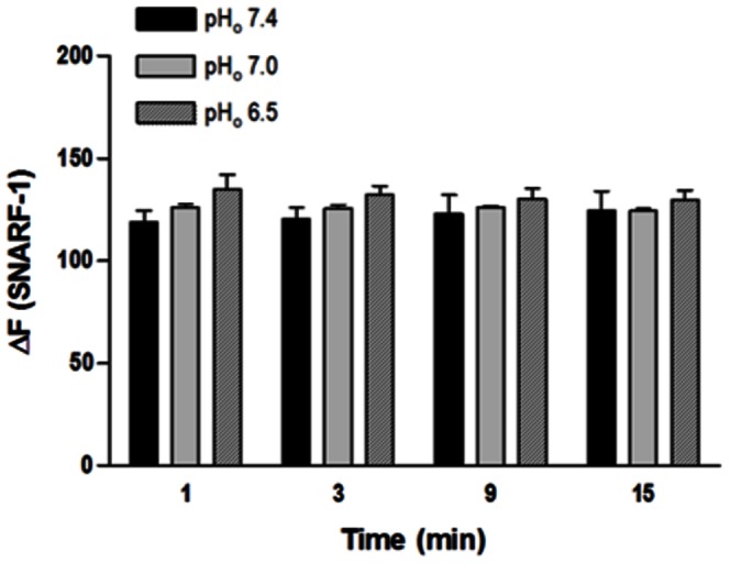
Figure 4. Effect of extracellular acidification on pHi in isolated endothelial cells from rat aorta.

Cells were loaded with SNARF-1 (10 µM) and analyzed by flow cytometry. HCl was used to reduce pHo from 7.4 to 7.0 and from 7.4 to 6.5; Hanks solution pH 7.4 served as control. Fluorescence intensity was measured before the HCl stimulus (F0) and at different time points (t = 1, 3, 9 and 15 min) after this stimulus (Ft). Results are reported as ΔF = Ft - F0. All values are means ± SEM (n = 7). Two-way ANOVA, Bonferroni's post-test.
