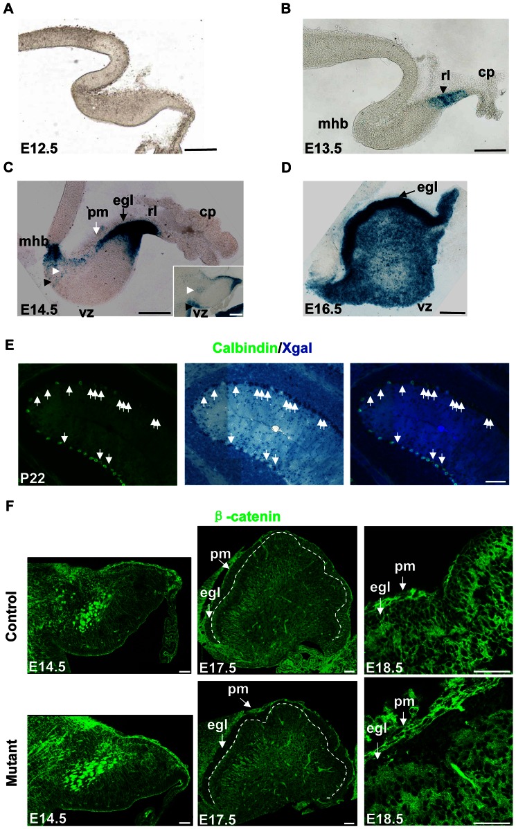
Figure 1. hGFAP-Cre activity in the cerebellum.
Sagittal sections were shown. (A) Xgal staining in an E12.5 hGFAP-Cre, R26R mouse, showing the absence of Cre activity in the cerebellum. (B) Xgal staining of an E13.5 hGFAP-Cre, R26R, β-cateninfl/fl mouse. The rl (black arrowhead) showed Xgal labeling. (C) Xgal staining of an E14.5 hGFAP-Cre, R26R, β-cateninfl/wt mouse. The rl and egl showed extensive Xgal labeling. Xgal labeling also appeared in the mhb, vz (black arrowhead) and the interior of cerebellum (white arrowhead). The inset is a lateral section from the same mouse, showing more staining in the vz region. There is no staining in the pm (white arrow). (D) Xgal staining of an E16.5 hGFAP-Cre, R26R, β-cateninfl/wt mouse. The egl (arrow) and the interior of the cerebellum showed extensive Cre activity. (E) A P22 hGFAP-Cre, R26R mouse first stained with Xgal, and then stained with calbindin antibody using a fluorescent secondary antibody. Some cells were double-positive (arrows) and formed Xgal clusters. (F) Sections from E14.5, E17.5, and E18.5 hGFAP-Cre, β-cateninfl/fl mice and control littermates stained with anti-β-catenin antibody. The level of β-catenin was normal in the E14.5 mutant mouse, but clearly decreased in the EGL of E17.5 or E18.5 mutant mice. The white dashed line in E17.5 sections indicates the inner border of the EGL. β-catenin was still expressed in the pm of mutant mice. Abbreviations: cp, choroid plexus; egl, external granule cell layer; mhb, mid-hindbrain boundary; pm, pia mater; rl, rhombic lip; vz, ventricular zone. Scale bar: 100 μm (A–E) and 50 μm (F).

