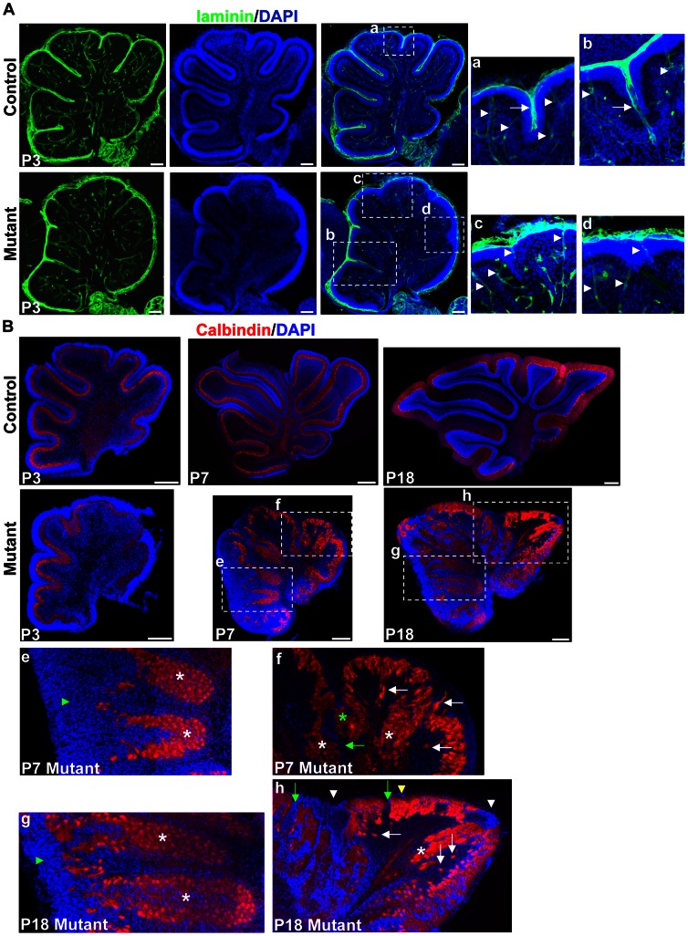
Figure 4. Lamination defects in the hGFAP-Cre, β-cateninfl/fl mouse.
(A) Sagittal sections through the cerebellar cortex of P3 mice stained with antibody against laminin and with DAPI. The right panels are enlargements of the squares in the middle panels. Note that laminin was deposited around blood vessels in control and mutant mice (arrowheads). In control mice, laminin was incorporated into the meningeal basement membranes overlying the EGL, and penetrated into the folia at fissures (arrow in a). In mutant mice, laminin either crossed the base of granule cell accumulation (arrow in b), or did not penetrate into the granule cell accumulation (c and d). (B) Midsagittal sections stained for calbindin and DAPI. At P3, mutant cerebella had a relatively normal PCL and EGL. The PCL was disrupted at P7. The squares are enlarged in the lower panels (e, f, g and h). Green arrowheads in e and g indicate deeply-located PCL covered externally by granule cells. In f and h, white asterisks indicate fused sites of the PCL from adjacent lobules. The green asterisk indicates the PCL fused in the middle of an unsplit fissure. White arrows indicate delaminated Purkinje cells, and green arrows indicate gaps in the PCL. White arrowheads indicate granule cell ectopia at the fusion lines between adjacent lobules, and yellow arrowhead indicates granule cell ectopia below the pial surface. Scale bars: 100 μm in (A) and 250 μm in (B).

