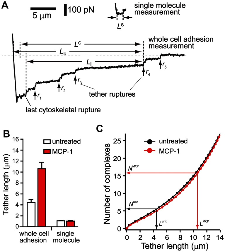
Figure 5. Individual tethers in whole cell measurements were supported by multiple α4β1/VCAM-1 complexes.
(A) Comparison of AFM force measurements acquired with extensive (whole cell measurement) and minimal (single molecule measurement) cell-substrate contact. L S is the length of tethers supported by a single α4β1/VCAM-1 complex as detected in single molecule measurements. In whole cell adhesion measurement shown, the cell detachment process involved the formation and breakage of at least 5 membrane tethers, labeled r 1 through r 5. Each tether is suppoted by multiple α4β1/VCAM-1 complexes. The lower and upper limits (L l and L u), and the average length (L C) of fourth tether (r 4) are shown. (B) Comparison of average tether lengths from whole cell measurements (L C) and tether lengths from single molecule measurements (L S). (C) Estimates of the number of α4β1/VCAM-1 complexes in a tether generated during the whole cell measurements. Black and red plots provide the range for the number of α4β1/VCAM-1 complexes in a single tether for untreated and MCP-1 stimulated cells, respectively. The superscripts unt and MCP associated with N (number of complexes) and L (tether length) designate untreated and MCP-1-stimulated cells, respectively.

