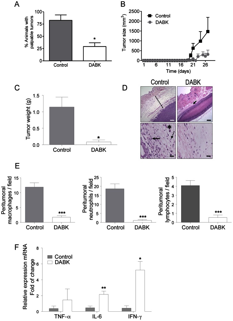
Figure 3. Stimulation of melanoma cells with the B1 receptor agonist reduces tumor growth and peritumor inflammatory infiltration after in vivo implantation.
(A) Incidence of animals with palpable tumor 28 days after injection of DABK stimulated cells. (B) Tumor growth curve from control and DABK-stimulated cells injected in C57/Bl6 mice. (C) Average tumor weight 28 days after tumor cell injection (D) Representative images at low magnification showing the size of the peritumor inflammatory infiltrate (upper panel, dashed line and arrow) and at high magnification showing the immune cells present in the tumor stroma (lower panel, * macrophage, arrow – neutrophil, dashed arrow – lymphocyte). (E) Peritumor inflammatory infiltrate assessed by quantification of the number of macrophages, neutrophils and lymphocytes in ten different high magnification fields (A = 400x; n = 6). (F) Detection of TNF-α, IL-6 and IFN-γ cytokine expression in the tumor mass as assessed by RT-qPCR (n = 6). In vivo studies of primary tumor growth n = 12; Data are expressed as the mean ± SEM; * p<0.05; ** p<0.01; *** p<0.001; DABK: desArg9-bradykinin; DLBK: desArg9-[Leu8]-bradykinin. Scale bars represent 200 μm and 50 μm in the upper and lower panels, respectively.

