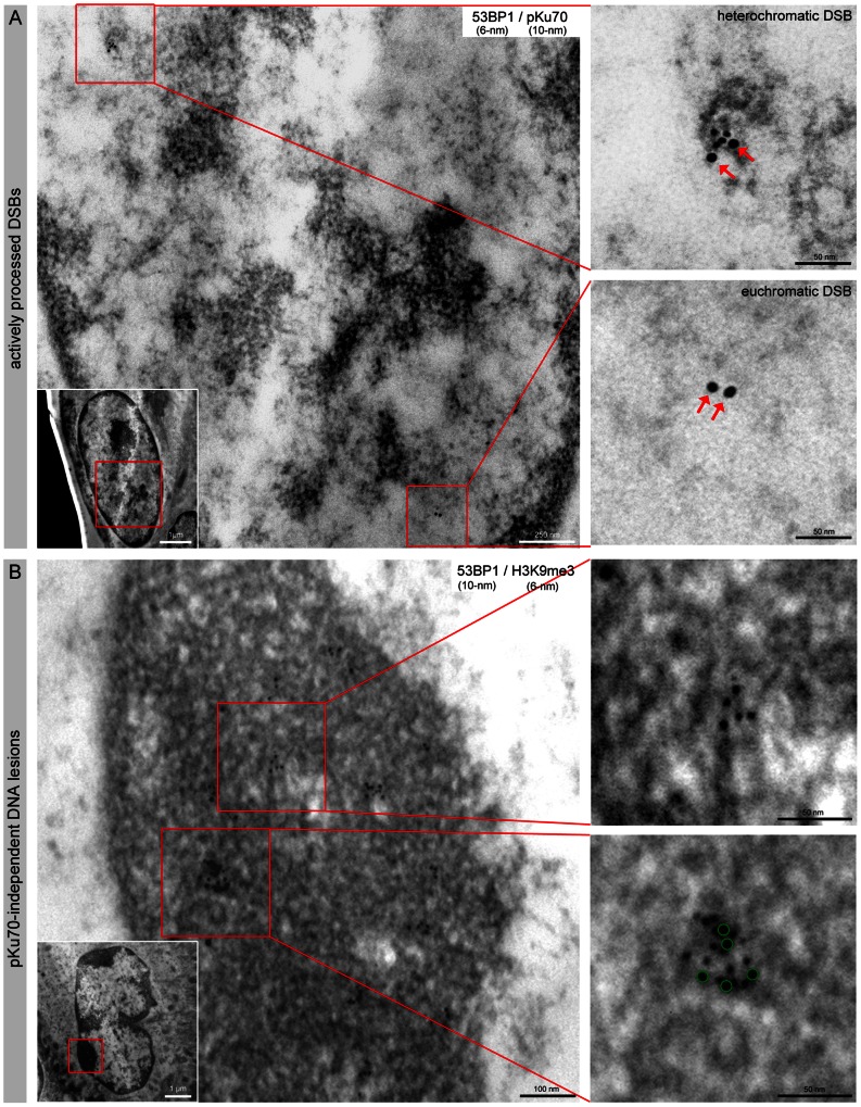
Figure 4. Characterization of radiation-induced DNA lesions by TEM.
(A) TEM micrographs of double-labeling of 53BP1 (6-nm) and pKu70 (10-nm) in HFSCs analyzed 0.5 h after 2 Gy irradiation. pKu70 consistently colocalized with 53BP1 in heterochromatic regions, but only pKu70 clusters (without 53BP1) were detected in euchromatic regions. (B) TEM micrographs of double-labeling of 53BP1 (10-nm) and H3K9me3 (6-nm) in HFSCs analyzed 72 h after protracted low-dose radiation (40× 10 mGy). Persistent 53BP1 clusters (green circles at higher magnification) colocalized with the heterochromatin marker H3K9me3 and were localized predominantly in tightly packed heterochromatin (dark grey regions in TEM; compare inset with the overview of the whole nucleus).

