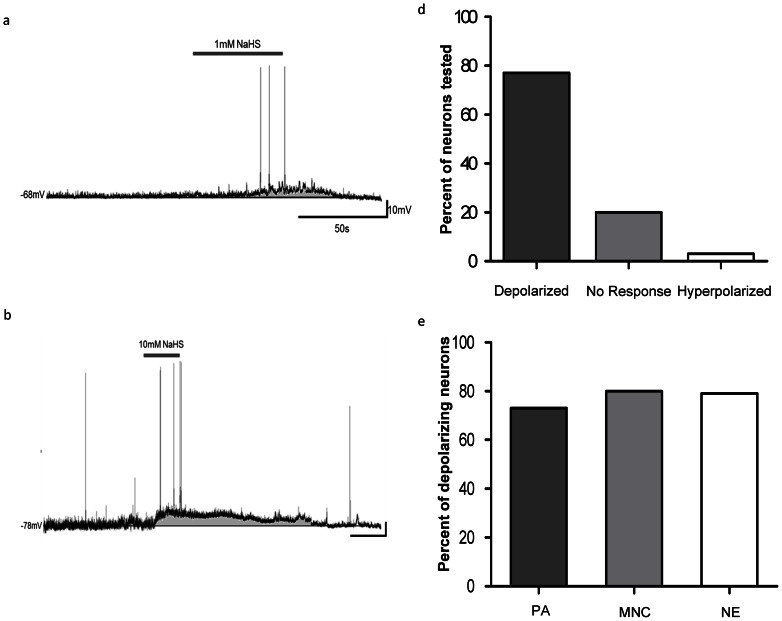
Figure 1. Hydrogen sulfide depolarizes PVN neurons.
Traces illustrate the depolarizing effects of NaHS application on PVN neurons. a) Current clamp recording trace illustrating a depolarizing response to 1 mM NaHS. Trace b) shows a current clamp recording illustrating a depolarizing response to 10 mM NaHS. Bar graph c) illustrates the various responses to NaHS (0.1–50 mM) of PVN neurons (80%, n = 52/65 depolarized, 20%, n = 13/65 showed no response, and 3%, n = 2/65 hyperpolarized). Bar graph d) shows the percentage of PA (73%, n = 24/33), MNC (80%, n = 12/15), and NE (79%, n = 11/14) neurons that depolarized.

