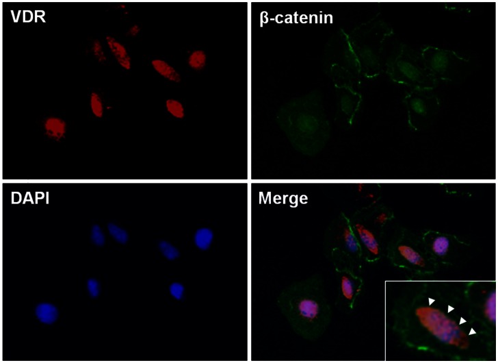
Figure 10. Nuclear co-localization of vitamin D receptor (VDR) and β-catenin in HK-2 cells.
Immunofluorescence staining demonstrated that the expression of VDR (red) was found to be co-localized with β-catenin (green) in the nuclei. Nucleus was stained with 4,6-diamidino-2-phenylindole (blue). The cells were pretreated with paricalcitol for 3 h, followed by 10 µM HHE treatment for 6 h (original magnification x 200). Inset show nuclear co-localization of VDR and β-catenin at a higher magnification indicated by arrow heads.

