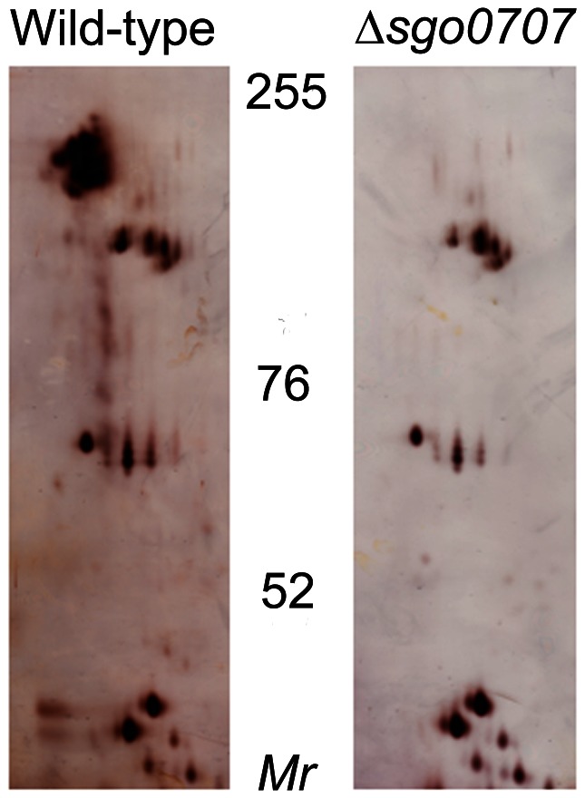
Figure 2. Silver-stained 2DE gels of cell wall proteins from wild-type and ΔSgo0707 strains of S.gordonii.
Proteins prepared as described in materials and methods were subjected to isoelectric focussing at pH 4–7 followed by SDS-PAGE in 7% gels. The migration positions of molecular mass markers are shown.

