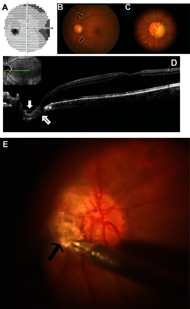
Figure 1.

Photographs of the left eye before pars plana vitrectomy (A–D) and during surgery (E). (A) Humphrey threshold 30–2 perimetry was performed at another hospital, and the images from that procedure show decreased sensitivity in the Bjerrum’s areas. (B and C) Fundus photographs demonstrating retinal elevation with folds at the macula and optic nerve head cupping with nerve fiber layer defects (arrows) but no obvious optic disc pit. (D) Optical coherence tomography image showing retinal schisis extending from the optic disc to the macula, without retinal detachment, as well as membrane tissue with a sheet-like shape (white arrow) on the optic disc and a tunnel-like hyporeflective lesion (black arrow) directly connecting the retinal schisis and the 4 o’clock margin of the disc. (E) The membrane tissue on the optic disc grasped by forceps.
