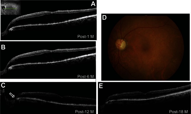
Figure 2.

Photographs of the left eye after vitrectomy. (A) Optical coherence tomography (OCT) image showing that the tunnel-like lesion in the optic disc was obscured 1 month after surgery and (B) retinal schisis was reduced 6 months after surgery. (C) Twelve months after surgery, OCT revealed that the tunnel-like hyporeflective lesion (black arrow) had resolved and retinal schisis had decreased further. (D) Fundus photograph taken 18 months after surgery showing the decrease in retinal elevation at the macula and no optic disc pit. (E) OCT image showing normalization of central retinal thickness with almost complete resolution of retinal schisis.
Abbreviation: M, month/s.
