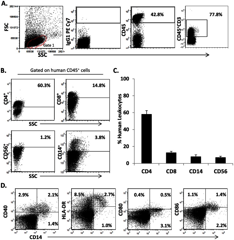
Figure 1. Production of the humanized BLT mouse to study TB.
NOD/SCID/γc null (NSG) mice were engrafted with human fetal liver and thymus, and supplemented with CD34+ cells. Shown in A, flow cytometry analysis displaying side scatter (SSC) and forward scatter (FSC) characteristics (Gate 1) of isolated peripheral blood from a representative BLT mouse twelve weeks post-engraftment. Plots 2–4 are the gating strategy for selection of cells expressing human CD45 pan leukocyte marker, the corresponding isotype control (IgG1 PE Cy7), and the CD3+ population subgate. B, percentage of the gated cells expressing markers for T cell subsets (CD4, CD8), NK cells (CD3−CD56+) and monocyte/macrophages (CD14+). C, average leukocyte % among gated CD45 cells in four groups of reconstituted BLT mice (n = 44) used for the subsequent studies (Fig. 2–8). D shows the expression of antigen presenting cells (APC) markers relevant to antigen presentation (HLA-DR) and T cell activation (CD40, CD80, CD86) expressed by peripheral blood monocyte/macrophages.

