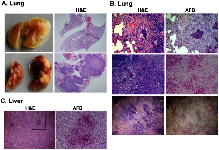
Figure 5. TB in humanized mice infected with a low dose of M.tb .
Animals were infected i.n. with 250 CFU tdTomato H37RV M.tb after verification of appropriate reconstitution with human leukocytes. Shown are images captured by brightfield microscopy following analysis of lung and liver from a representative animal (n = 11). The images demonstrate tissue damage and inflammation (hematoxylin and eosin, H&E) localized to M.tb bacilli (acid fast bacilli, AFB) in formalin-fixed tissue sections from lung and liver of BLT mice sacrificed at 6–8 weeks p.i. Shown in A are gross lung lobes (left panels) and cross sections of whole lung stained with H&E, captured using a stereomicroscope. B, Lung tissue pathology visualized by H&E staining (left panels) and AFB (right panels). Top panels (20X) demonstrate bronchial obstruction in the lung and the large numbers of bacteria within the obstruction. Middle panels (20X) show cholesterol crystal deposits (black arrow) observed in large granulomas at later stages (≥6 wk) of infection. Bottom panels (4X) shows center of large, coalescing granulomas characterized by necrosis and lack of AFB. C, shown is liver tissue pathology (left panel, 10X) and AFB (right panel, 40X) in the indicated region). Images are from lung and liver of a representative animal (n = 6).

