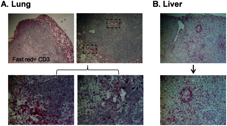
Figure 6. Human T cells are recruited to and organize at lung granulomas and sites of inflammation following M.tb infection.
Animals were infected i.n. with 250 colony forming units (CFU) tdTomato H37RV M.tb following establishment of human immune cell populations. Formalin fixed paraffin embedded tissue sections were cut, dewaxed and stained with antibody to human CD3. Marker expression was visualized with Fast Red substrate and images captured by brightfield microscopy. A, shown is the localization of T cells relative to a granuloma periphery and center (top left; 4X, top right; 10X). Enlarged images of T cell staining in the indicated areas are shown in the bottom panels (40X). (B) Human T cells in portal tracts and sites of inflammation in the liver (top panel, 10X) and an enlarged area showing orchestration of T cells around an inflammatory focus (bottom panel, 20X). Shown are representative images (n = 3) of animals sacrificed at 7 wk p.i.

