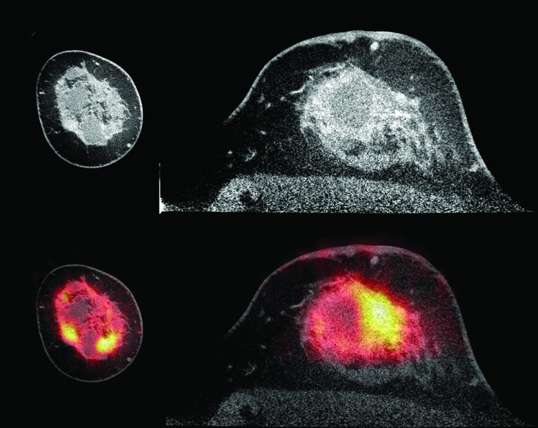
Figure 8.
Images of a patient with an invasive ductal carcinoma with lobular features obtained using a dedicated LYSO PET/CT system. The contrast enhanced CT, shown at top (coronal view at left and axial view at right), has been window to enhance appearance of benign cysts. The fused PET/CT image, at bottom, demonstrates F-18 FDG uptake in the extensive tumor. Image courtesy of Dr. Ramsey Badawi, University of California Davis.

