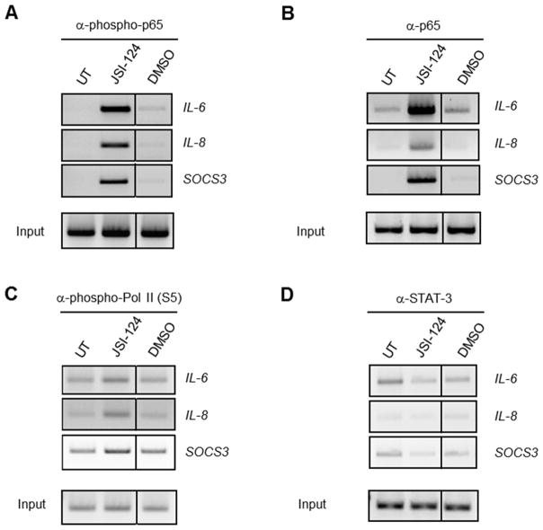
Figure 4. ChIP reveals the presence of phosphorylated p65 and RNA Pol II at the promoters of IL-6, IL-8 and SOCS3 of JSI-124 treated cells.
U251-MG cells were left untreated (UT) or treated with JSI-124 (1 μM) or DMSO (vehicle) for 1 h. Immunoprecipitation was performed with 5 μg of Ab to phospho-p65 (A), total p65 (B), phospho-RNA Pol II (C) and STAT3 (D). The immune complexes were absorbed with protein A beads or protein G beads blocked with bovine serum albumin and salmon sperm DNA. Immunoprecipitated DNA was then subjected to semi-quantitative PCR and analyzed by gel electrophoresis for IL-6, IL-8 and SOCS3. The experiment was repeated, and similar results were observed.

