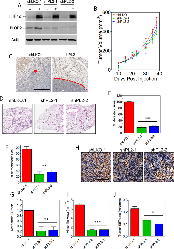
Figure 6.
PLOD2 is essential for invasion and metastasis of MDA-MB-435 cells. A, immunoblot assays were performed using lysates prepared from control (shLKO.1) or PLOD2-knockdown (shPL2-1 and shPL2-2) MDA-MB-435 subclones exposed to 20% or 1% O2 for 48 hours. B, the indicated subclones were injected into the MFP of NOD-SCID mice and tumor volume was plotted versus time. C, hematoxylin staining of primary tumor sections. Invasion of shLKO.1 into adipose tissue (red arrow) is shown in the left panel. The well-defined boundary between shPL2 cells and normal tissue is indicated by the dashed line in the right panel. Scale bar = .5 mm. D, lung sections (5×5 mm) were stained with hematoxylin and eosin. E, metastatic area was determined by image analysis (mean ± SEM, n = 5, one-way ANOVA). F, the number of metastatic foci per 5×5 mm tumor section was determined (mean ± SEM, n = 5, one-way ANOVA). G, human genomic DNA content in mouse lungs was quantified using qPCR with human-specific HK2 gene primers (mean ± SEM, n = 5, one-way ANOVA). H, ipsilateral axillary lymph node sections were subjected to immunohistochemistry using an antibody specific for human vimentin. Scale bar = 100 µm. I, vimentin staining was quantified by image analysis (mean ± SEM, n = 5, one-way ANOVA). J, the stiffness of freshly dissected control or shPLOD2 tumors was determined. Bonferroni post-tests were performed for all ANOVAs. *P<0.05, **P < 0.01, ***P < 0.001 vs. shLKO.1.

