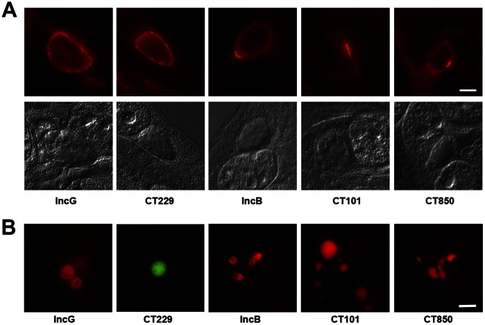
Figure 1. Inclusion membrane localization of specific Incs and corresponding structures when ectopically expressed.
A. C. trachomatis L2 inclusions at 18 hr post-infection stained for immunofluorescence with specific antibodies to the inclusion membrane proteins IncG, CT229, IncB, CT101, and CT850. IncG and CT229 show circumferential staining patterns while IncB, CT101, and CT850 are enriched in microdomains on the inclusion membrane. Nomarski differential interference contrast images of the same fields are shown for reference. B. The same Incs as above ectopically expressed in HeLa cells as mCherry or GFP fusions. Bar = 10 µm.

