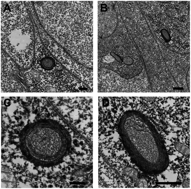
Figure 2. Immunoelectron microscopy of ectopically expressed mCherry-IncB in HeLa cells.
A and B. Examples of mCherry-IncB expressed in HeLa cells and immunolabled with an anti-mCherry antibody followed by an HRP-conjugated secondary antibody and developed with a commercial diaminobenzidine substrate. C and D. Higher magnification of the same sections showing internal membrane structure. Bars = 1 µm (A&B); 0.5 µm (C&D).

