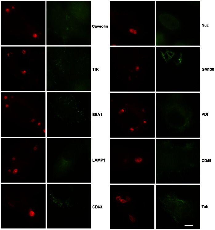
Figure 5. Immunofluorescent staining for various cellular organelles and structures in mCherry-IncB expressing HeLa cells.
No colocalization of any markers (green) with IncB induced vesicles (red) was observed. Markers included: Caveolin, transferrin receptor (TfR), early endosomal antigen 1 (EEA1), lysosomal glycoprotein 1 (Lamp1), CD63, nucleolin (Nuc), GM130, protein disulfide isomerase (PDI), CD49, and tubulin (tub). Bar = 10 µm.

