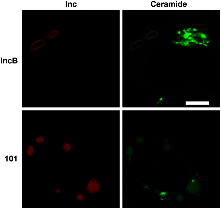
Figure 8. Labeling of CT101 and IncB induced vesicles with C6-NBD-ceramide.
Cultures were visualized after 5 min of back-exchange [40], [41] to extract plasma membrane localized fluor. Note the distinct rim-like staining patterns of the IncB-induced vesicles as compared to the internal structure of the CT101-induced vesicles. The intense green staining by C6-NBD-ceramide represents the Golgi apparatus. Bar = 10 µm.

