Fig. 1.
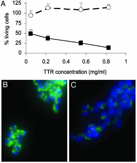
(A) Cytotoxicity of WT TTR (○) and V30M TTR (▪) on IMR-32 cells after 3 days of incubation at 37°C. Bars represent the SD of triplicate determinations. (B and C) Fluorescent microscopy images of IMR-32 cells stained with Rhodamine 123 (green) and Hoechst 33342 (blue) after 40 h incubation with 1 mg/ml V30M TTR. (B) Control cells (Opti-MEM only). (C) V30M TTR-treated cells. The decrease in mitochondrial membrane potential (green) of TTR-treated cells compared with control cells, suggest that the TTR triggers apoptosis in IMR-32 cells. Some nuclei condensation (clear blue) is seen also after TTR treatment.

