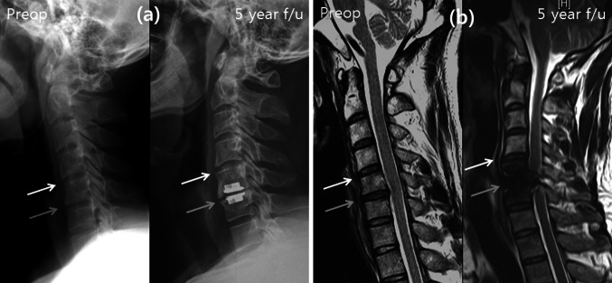
Fig. 3.
Radiography and MRI of arthroplasty patient at preoperative and 5-year follow-up. Grey arrow indicates operation site, and white arrow indicates upper adjacent segment. At X-ray, there was no significant disc space narrowing and posterior osteophytes (a), but there was a disc herniation and signal change at upper adjacent segment at MRI (b)

