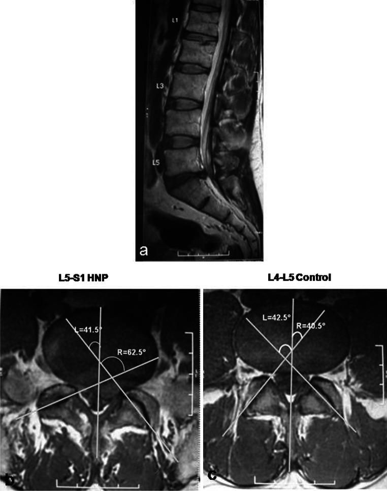
Fig. 2.
a 39-year-old patient had a left sided disc herniation at L5–S1. The L4–L5 disc served as control. b The axial image shows the presence of facet tropism at the level of disc herniation, with the disc herniating towards the side of the sagittally-oriented facet. c The axial cut at the level of the L4–L5 disc (control) shows that the facets are symmetrical

