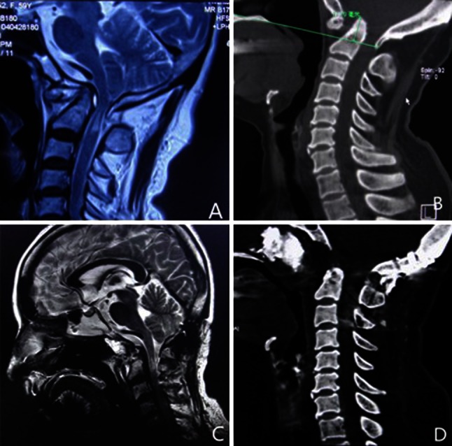
Fig. 3.

Case 3: a Preoperative magnetic resonance imaging (MRI) revealing basilar invagination, resulting in compression at the cervicomedullary junction from the ventral aspect. b Preoperative sagittal computed tomographic (CT) scans revealing the tip of the odontoid extended above the Chamberlain line 17.2 mm. c Two weeks postoperative, MRI showed the ventral cervicomedullary compression was completely relieved. d Two days postoperative, sagittal computed tomographic (CT) scans revealing that the odontoid had been drilled off
