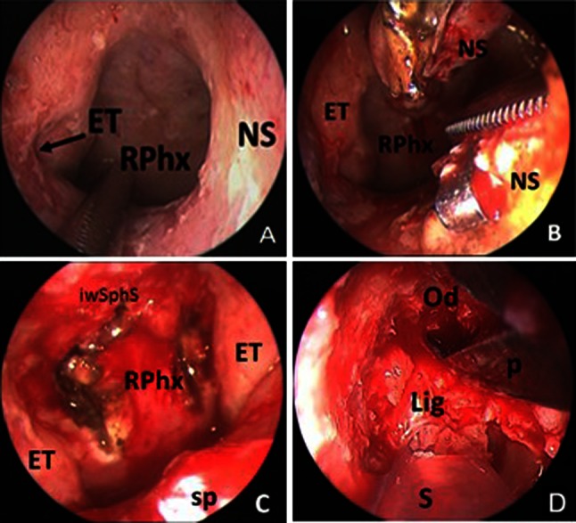
Fig. 5.

a The choana was enlarged although the bilateral middle turbinates, inferior turbinates and the sphenoid sinus anterior wall were not resected. b The posterior 1 cm of the nasal septum was removed to enlarge the choana to facilitate bilateral application of instrumentation. c The fascia of the posterior nasopharynx was opened with an inverted U incision. The superior extent is at the clivus and the lateral margins just medial to the Eustachian tubes. d The ligaments and dura were seen after most of the odontoid was removed (ET Eustachian tube, iwsphs inferior wall of the sphenoid sinus, NS Nasal septum, RPhx rhinopharynx, sp soft plate, Od odontoid, Lig ligaments, S suction, P punche)
