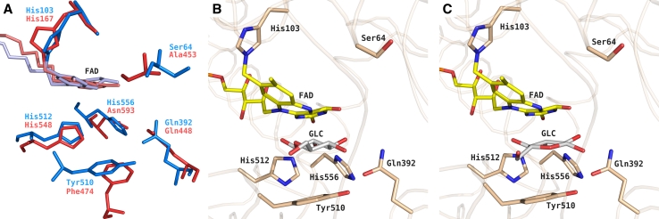
Fig. 1.
Active site structures of homotetrameric pyranose 2-oxidase (P2O) from Trametes multicolor and monomeric pyranose dehydrogenase (PDH) from Agaricus meleagris. a Superposition of active site residues from PDH, blue (PDB: 4H7U) and P2O, red (PDB: 3LSK). For clarity, the FAD moiety was colored light blue in PDH or light red in P2O and the atoms of the FAD ribityl side chains were omitted. b, c After superpositioning, d-glucose coordinates were grafted from the P2O structure with PDB accession codes (b) 3PL8 or (c) 2IGO into PDH. For clarity, P2O is not shown. Atom-coloring scheme: carbon (beige, protein; yellow, FAD; white, ligand), nitrogen (blue), oxygen (red), and phosphate (orange). Figures were generated using PyMOL (http://www.pymol.org/)

