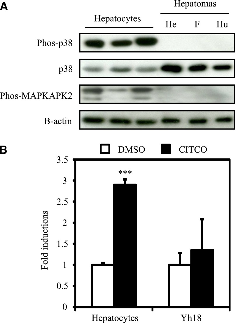
Fig. 1.
Phosphorylation of p38 MAPK and CAR-mediated activation of CYP2B6 in human primary hepatocytes and hepatoma cell lines. (A) Hepatocyte and cell extracts were prepared as described in Materials and Methods from seeded hepatocytes and cells without any exposure. Protein levels of phosphorylated p38 MAPK (Phos-p38), p38 MAPK (p38), and phosphorylated MAPKAPK2 (Phos-MAPKAPK2). Protein levels of B-actin were determined as an internal control. Data shown are three individual donors of primary hepatocytes and three different hepatoma cell-lines (HepG2, FLC7, and Huh7 cells from left to right). He, HepG2 cells; F, FLC7 cells; Hu, Huh7 cells. (B) Total RNAs were prepared as described in Materials and Methods from one female donor of hepatocytes and Yh18 cells treated as described in Materials and Methods. Expression level of CYP2B6 mRNA was determined. Values are expressed as fold inductions relative to that of CYP2B6 mRNA level normalized to the expression levels of B-actin (ACTB) mRNA in dimethylsulfoxide-treated hepatocytes or Yh18 cells. Data are mean ± S.D. (n = 3 in each group). ***P < 0.005 for comparison between with and without CITCO exposure, Newman-Keuls multiple comparison test.

