Fig. 2.
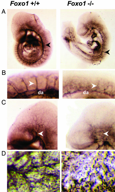
Foxo1-/- embryos and yolk sacs show defective vascular development. (A) PECAM-1 immunostaining of whole-mount E9.5 Foxo1+/+ and Foxo1-/- embryos, respectively. Note the thin and disorganized dorsal aorta in Foxo1-/- embryo compared with Foxo1+/+ embryo (arrowheads). (B) Magnified view of the E9.5 Foxo1+/+ and Foxo1-/- intersomitic vessels. Intersomitic vessels in Foxo1-/- embryo were disorganized compared with Foxo1+/+ (arrowheads). da, dorsal aorta. (C) Magnified view of the E9.5 Foxo1+/+ and Foxo1-/- head vessels. The vessels of the head, including the branches of the internal carotid artery, were properly developed in Foxo1+/+ but not in Foxo1-/- embryos (arrowheads). (D) PECAM-1 staining of Foxo1+/+ and Foxo1-/- yolk sacs. Properly developed vasculature was present in Foxo1+/+ yolk sacs but not in Foxo1-/- yolk sacs.

