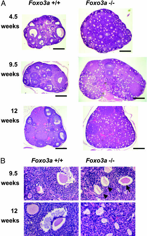
Fig. 5.
Histological analysis of ovaries with hematoxylin/eosin staining. (A) At 4.5 weeks of age, developing follicles containing growing oocytes but no antrums (type 3b-5b) were prominent, but mature follicles containing antrums (type 6-7) were less common in Foxo3a-/- ovaries. At 9.5 weeks of age, Foxo3a-/- ovaries had many developing follicles and few mature follicles, similar to those at 4.5 weeks. At 12 weeks of age, Foxo3a-/- ovaries had no developing follicles, and all oocytes had undergone degeneration. Each specimen was sectioned at its largest diameter. (Scale bar, 500 μm.) (B) Various stages of follicles were found in wild-type ovaries at both 9.5 and 12 weeks of age. Normal oocytes (arrow) were present, but degenerating oocytes (arrowheads) were prominent in Foxo3a-/- ovaries at 9.5 weeks of age. All oocytes had undergone degeneration in Foxo3a-/- ovaries at 12 weeks of age.

