Fig. 3.
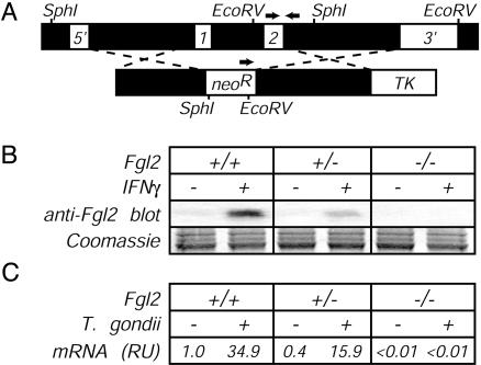
Production of Fgl2-deficient mice. (A Upper) Schematic of the Fgl2 gene, indicating (i) locations of the two Fgl2 exons, (ii) restriction enzyme sites and probes used for confirming successful disruption of the fgl2 gene by Southern blotting, and (iii) locations of PCR primers (arrows) used to routinely screen for the targeted mutation. (Lower) Schematic of the targeting construct, indicating regions flanking the fgl2 exons that were cloned into the targeting vector and the relative placement of selectable elements. neoR, neomycin resistance gene; TK, thymidine kinase gene. (B) Impaired secretion of Fgl2 protein by Fgl2-deficient cells. Thioglycollate-elicited peritoneal macrophages were isolated from littermate mice of the indicated fgl2 genotype. After 24 h of culture in the presence or absence of IFNγ (20 ng/ml), cell-free supernatants were assayed for Fgl2 expression by Western blotting with Fgl2-specific mAb. Coomassie blue staining of a gel run in parallel documented equivalent sample loading in all lanes. (C) Impaired production of Fgl2 mRNA by T. gondii-infected Fgl2-deficient mice. As in Fig. 2, mice were infected with T. gondii, liver tissue was harvested on day 8 after infection, and levels of Fgl2 mRNA were determined by real-time PCR. Data depict the average of four to five mice per group after normalization to uninfected WT mice.

