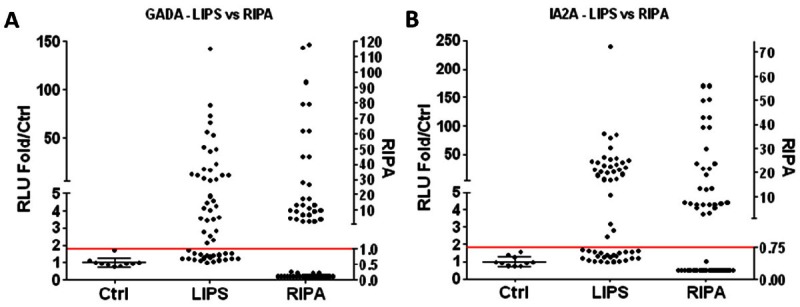
Figure 1.

Detection of GADA and IA-2A by LIPS. Sera (n = 54) that were tested for ICA, GADA, and IA-2A clinically by RIPA or healthy normal donor control sera (n = 10) were used in panels A, B, and the transverse line is 3SD above control mean. RLU (relative light units) are expressed as fold relative to control mean (left y-axis). Standard LIPS assays (1μl serum) were performed with (A) Rluc-GAD65 fusion protein lysate or (B) Rluc-IA-2 fusion protein lysate and are compared to clinical RIPA data (right y-axis).
