
Figure 1.
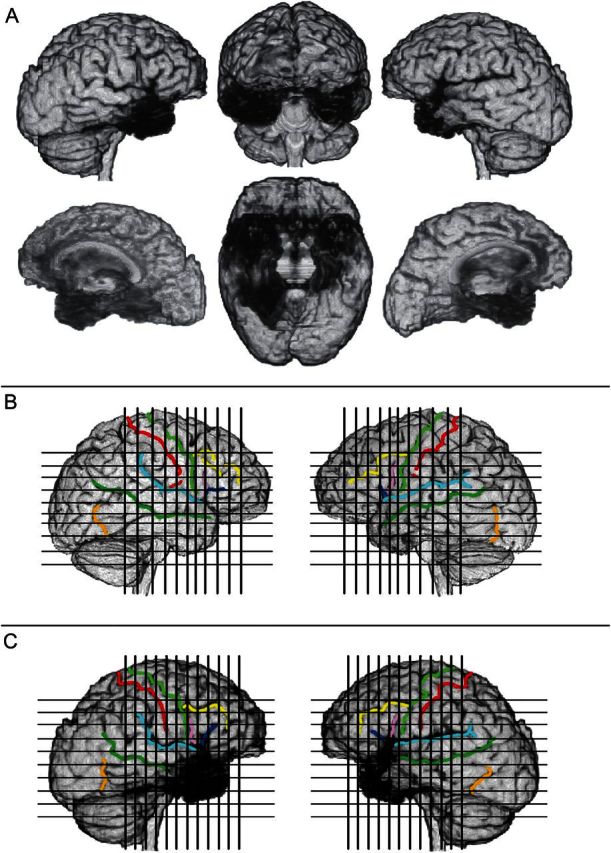
(A) Three-dimensional reconstruction of patient B’s brain, using Brainvox. The right hemisphere is shown on the left (lateral view on top; mesial view below). The same 2 views of the left hemisphere are depicted on the right. The middle column shows the brain seen from the front (top) and a ventral view of the 2 hemispheres after removal of the cerebellum and brainstem (bottom). The black shaded areas reveal the extensive damage involving a large sector of both temporal lobes, the posterior aspect of the orbital frontal region, and the anterior cingulate. (See Figs 2–4 for details). (B and C) Markings of major sulci (central sulcus = red; precentral sulcus = light green; inferior frontal sulcus = yellow; horizontal branch of the Sylvian fissure = dark blue; ascending branch of the Sylvian fissure = pink; Sylvian fissure = light blue; superior temporal sulcus = dark green; anterior occipital sulcus = light brown); and positioning of coronal and axial slices as shown in Figures 2 and 3, in the brain of a normal subject, the comparison brain (B), and in Patient B. (C) The comparison brain was obtained in the same scanner used for Patient B.