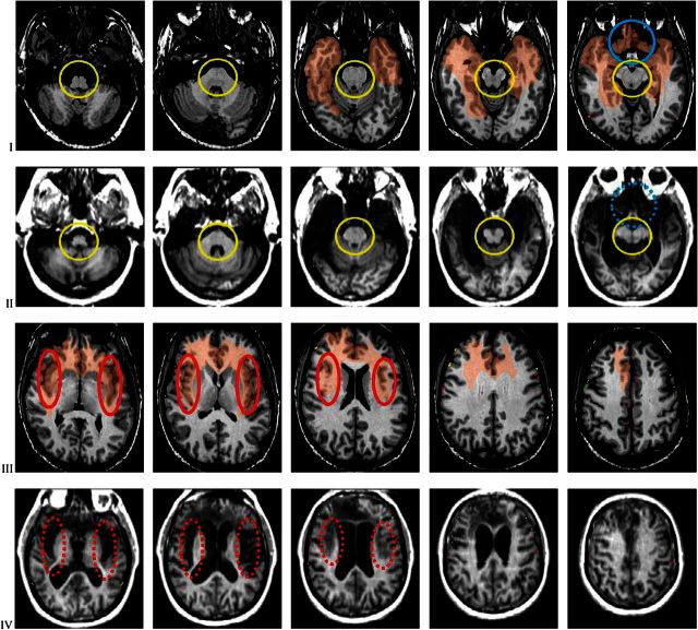
Figure 3.
Axial slices of the comparison brain and of patient B’s brain, interleaved as in Figure 2. The comparison brain shows the transfer of the area damaged in Patient B. Slice setting as in Figure 1B,C. There is again evidence of the complete damage of the insula, the underlying white matter and the claustrum in both hemispheres, highlighted by the red ovals. See panels 1–3 in rows III and IV. The damage also encompasses both temporal lobes destroying the polar regions, the amygdalae, the hippocampuses, and the parahippocampal gyri (panels 3–5 in rows I and II). In the right hemisphere, the damage extends further posteriorly in the inferior and mesial aspects. The damage involves the posterior fronto-orbital region bilaterally (highlighted by the blue circles in panel 5, rows I and II); the damage extends dorsally to involve the anterior cingulate cortex subcalosally and beyond (panel 1 in rows III and IV), and the white matter in the core of both frontal lobes more extensively in the right hemisphere (all panels in rows III and IV). The brainstem and cerebellum are intact (see panels 1–5 in rows I and II; brainstem circled in yellow), as are all sensory and motor cortices, and the association cortices of the parietal and occipital lobes, and the primary visual regions.

