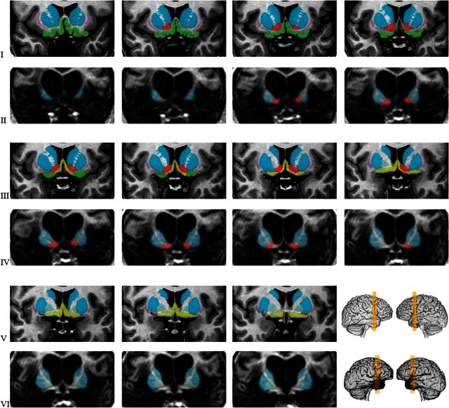
Figure 4.
Detail of the posterior orbital sector, basal forebrain region, and basal ganglia in the comparison brain and in patient B. The yellow colored slabs in the right lower corner represent the section of brain from which the panels of this figure were extracted. The slices in patient B. are 1.5-mm thick, and all consecutive cuts are shown. The slices in the comparison brain are anatomically matched to those of patient B. Rows of panels from the comparison brain are interleaved with those from Patient B., as in the previous figures. Several structures are colored. The same color is used for the comparison brain and Patient B’s brain. In Patient B., the dorsal striatum (putamen and caudate nuclei, in blue) appears intact. The ventral striatum/nucleus accumbens region (red), largely located below and in front of the anterior commissure is also seen in patient B. The whole basal ganglia complex appears to be diminished in size; partial damage is possible (see text). The basal forebrain nuclear masses (septal nuclei; diagonal band; substantia innominate), shown in greenish yellow in the comparison brain, are missing in Patient B. The posterior orbitofrontal cortices (green) and the claustrum (pink) are also missing in Patient B.

