Figure 2.

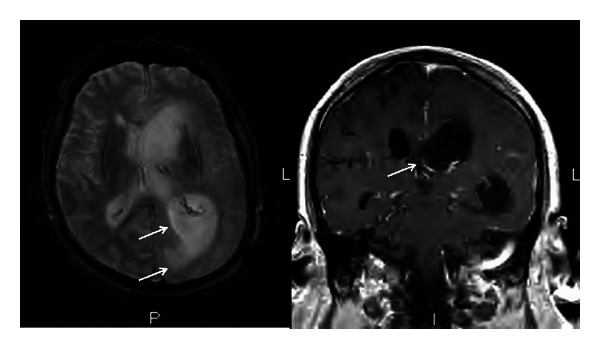
Brain MRI. Ventriculomegaly, increased halo surrounding the lateral ventricles, and debris within the ventricles with restricted diffusion and ependymal and subependymal enhancement (arrows).

Brain MRI. Ventriculomegaly, increased halo surrounding the lateral ventricles, and debris within the ventricles with restricted diffusion and ependymal and subependymal enhancement (arrows).