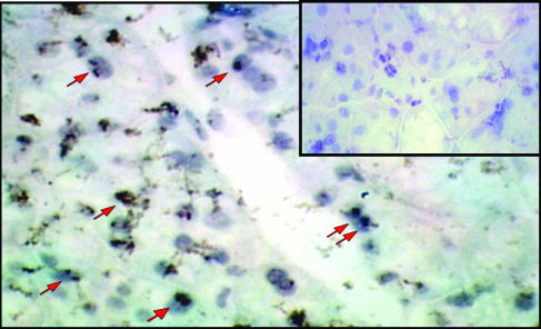
Fig. 6.
Immunocytochemical detection of β-galactosidase expression in mouse submandibular glands. Cryosections were prepared 8 weeks after AAVLacZ administration to mouse submandibular SGs (n = 5, 109 particles per animal), and β-galactosidase expression was detected by using an anti-β-galactosidase antibody. Sections were counterstained with hematoxylin. The β-galactosidase cDNA used contains a nuclear localization signal. Red arrows indicate representative cells with nuclear localized β-galactosidase. Staining was observed only in salivary ductal cells. No staining was detected in the control cryosections obtained from an animal receiving AAVhEPO by using the same immunocytochemistry procedure, as shown (Inset).

