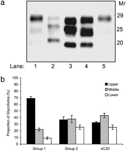
Fig. 4.
Electrophoretic analysis of PrPSc in cattle TSE and sCJD. (a) Western blot detection of PrPSc in brains of group 1 animals (lanes 1 and 5); subject with sCJD and type 1 PrPSc, methionine/methionine at codon 129 (lane 2); subject with sCJD and type 2 PrPSc, methionine/valine at codon 129 (lane 3); and group 2 cattle (lane 4). (b) Relative proportions of the three PrPSc glycoforms in group 1 and group 2 cattle compared with glycoform profiles obtained in nine sCJD patients, methionine/valine at codon 129 and with type 2 PrPSc. Mean ± standard deviation is shown. Upper band, diglycosylated form; middle band, monoglycosylated form; and lower band, unglycosylated form.

