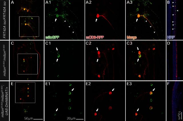
Figure 3.
milton33-853results in a loss of mitochondrial anterograde transport in larva motor neuron MARCM clones. Third-instar larval MN MARCM clones and their mitochondria were simultaneously labeled by expressing UAS-mCD8-RFP and UAS-mitoGFP under the MN-specific driver D42-Gal4 (D42-Gal4>UAS-mCD8-RFP, UAS-mitoGFP). HRP immunostaining was used to highlight far distal segmental nerves containing mCD8 RFP-expressing axons (B, D, F, ∼800 μm from the VNC). MitoGFP was distributed throughout the cell body (A, arrows), dendrites, as well as proximal (A, arrowheads) and distal axons (B) of wild-type MNs. In milton33-853MNs, mitoGFP signal was restricted to MN cell bodies (C, arrows) and absent from dendrites and axon (C and D). Overexpression of DmNMNAT (D42-Gal4>UAS-DmNMNAT) in milton MNs did not suppress the mitochondria trafficking defect, as mitoGFP was also restricted to cell bodies (E, arrows) and absent from dendrites and axons (E and F). Scale bars, 50 μm in (E) for (A), (C) and (E), 20 μm in E1 for A1–A3, C1–C3, and E1–E3, and 10 μm for (B), (D) and (F).

