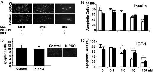
Fig. 3.
IR expression is required for insulin-stimulated inhibition of apoptosis in cultured neurons but not in the intact animal. (A) Cultured cerebellar granule cells of 6-day-old NIRKO (Upper) and control (Lower) mice were subjected to low KCl-containing (5 mM) medium to induce apoptosis (Left); cells were incubated with 100 nM insulin (Center) and with 100 nM IGF-1 (Right). Condensed nuclei were visualized by Hoechst dye 33342. (B) After reduction of KCl concentrations, cells were stimulated with 0, 0.1, 1.0, 10, and 100 nM insulin; control (open bars) and NIRKO (filled bars) mice. Data represent the mean ± SEM of at least eight animals of each genotype. (C) Antiapoptotic effect of IGF-1 on primary cerebellar granule cells. After reduction of KCl concentrations, cells were stimulated with 0, 0.1, 1.0, 10, and 100 nM IGF-1; control (open bars) and NIRKO mice (filled bars). Data represent the mean ± SEM of at least eight animals of each genotype. (D) Terminal deoxynucleotidyltransferase-mediated dUTP nick end labeling (TUNEL) assays of cerebella from postnatal-day-5 pups of NIRKO mice and their littermate controls. Quantification of apoptotic nuclei in the outer granule layer. Data represent the mean ± SEM of at least four animals of each genotype. (Magnification, ×40.)

