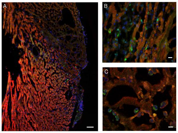
Figure 5.
Histological evaluation confirms BMMC homing to the infarcted heart. Fluorescence microscopy images of a representative heart 2 days following 30 minutes of I/R injury and injection of 5×10⁶ BMMN cells via tail vein. All panels stained with anti-troponin (red), anti-GFP (green), and DAPI (blue). (A) Infarcted area demonstrated by lack of bright troponin stain, with numerous GFP positive cells in the infarct border zone (scale bar = 50 μm). (B) High-power view demonstrating numerous GFP-expressing cells within the myocardium (scale bar = 10 μm). (C) Confocal laser microscopy image confirming presence of GFP-expressing cells within infarcted areas of the myocardium (scale bar = 5 μm).

