Figure 2.
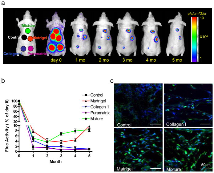
Bioluminescence imaging of transplanted MSCsFluc+/eGFP+ in living animals. (a) To assess longitudinal cell survival, animals were imaged for 5 months after subcutaneous injection of 5×10⁵ MSCsFluc+/eGFP+ mixed with PBS control, Matrigel, Collagen 1, Puramatrix, and mixture. (b) Quantification of BLI signals showed a drastic decrease of Fluc activities from day 2 to month 1. After that, the BLI signals in the Matrigel and mixture groups remained stable compared with the other three groups. BLI signals were all normalized to day 0 in each group. (c) Postmortem immunohistochemistry staining of eGFP by confocal fluorescence microscopy revealed more robust engraftment of MSCsFluc+/eGFP+ within the Matrigel and mixture groups compared to the PBS control and Collagen 1 groups, consistent with the noninvasive BLI data. Scale bars: 50 μm.

