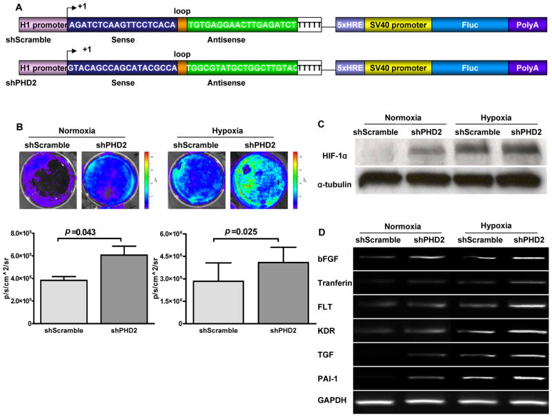
Figure 2. In vitro characterization of mouse shPHD2.
(a) Schema of classic hairpin carrying the site-2 sequence (shPHD2) and control hairpin carrying the scramble sequence (shScramble). The H1 promoter drives the expression of a hairpin structure in which the sense and antisense strands of the siRNA are connected by a 9-bp long loop sequence. In addition, a separate 5xHRE-SV40 promoter driving firefly luciferase (Fluc) is used to track shRNA activity in vitro and in vivo. 5x HRE, 5 repeat of hypoxia response elements; Sv40, simian virus 40. (b) In vitro imaging results indicate that Fluc signals increased significantly in respone to shPHD2 therapy during both normoxia and hypoxia conditions via binding of HIF-1α protein on the 5xHRE binding site. In addition, there were significant Fluc signal differences between shPHD2 and shScramble under normoxia (P=0.043) and hypoxia (P=0.025) states. (c) Similarly, Western blot data show that levels of HIF-1α protein were more robust after shPHD2 plasmid transfection during normoxia and and 6 hr hypoxia incubation. (d) RT-PCR analysis confirmed significant upregulation among 6 genes involved in angiogenesis due to activation of the HIF-1α protein from knocking down PHD2. GAPDH was used as the loading control.

