
Figure 5.
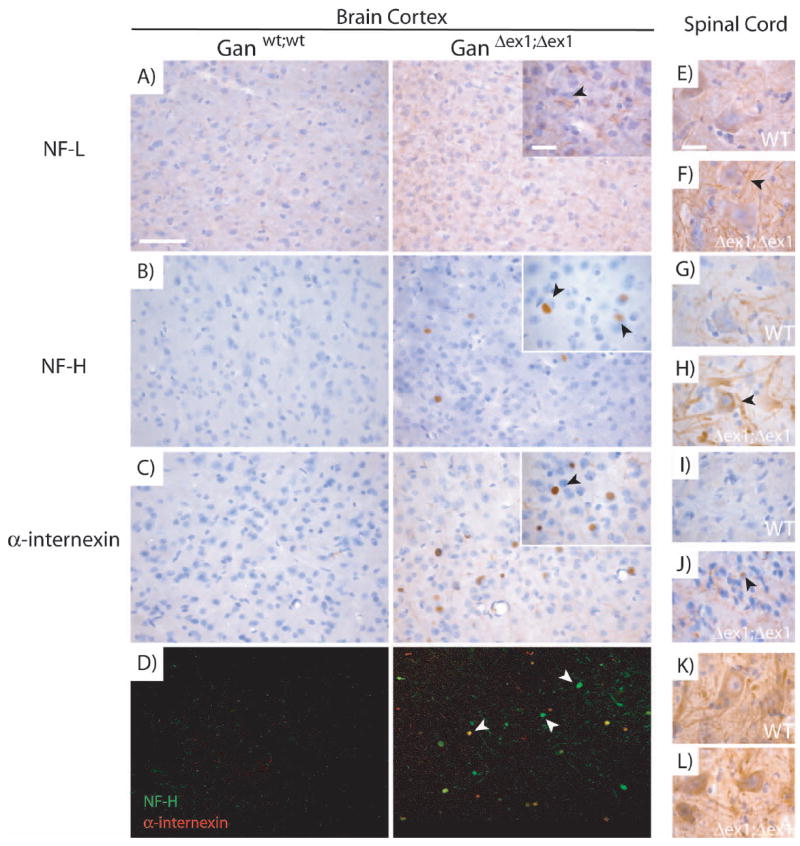
Accumulations of IFs in specific regions of the CNS in GanΔex1;Δex1 mice.
NF-L immunostaining was stronger in GanΔex1;Δex1 mice compared to normal mice (A). NF-H (B) and α-internexin (C) immunostaining revealed accumulations in the form of inclusion bodies in GanΔex1;Δex1 cerebral cortex. (D) Double labelling of NF-H and α-internexin in the cerebral cortex. NF-H and α-internexin do not always colocalize. NF-L (E-F) and NF-H (G-H) staining tend to be stronger in the lumbar spinal cord of motor neurons in GanΔex1;Δex1 mice unlike peripherin (K-L) which does not change. α-internexin (I-J) tends to form accumulations in the dorsal horn of the spinal cord as in the brain cortex. (Scale bar 100 and 10 μ.)