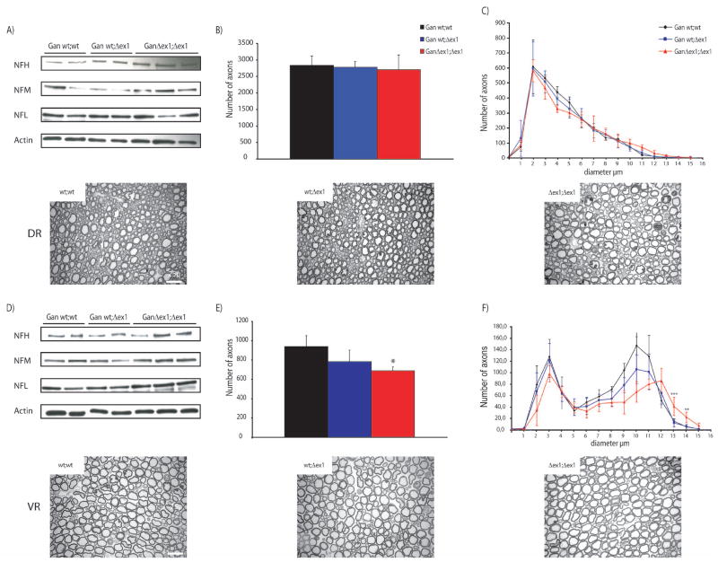
Figure 6.
The number of axons is lower than normal in GanΔex1;Δex1 L5 ventral root but not in dorsal root.
The content of NFs was reduced in GanΔex1;Δex1 L5 ventral root (D) but not in dorsal root (A). (B,E) show the average number of axons of wt;wt, Δex1;wt and Δex1; Δex1 mice in dorsal and ventral root, respectively. The number of axons was decreased by 27% in GanΔex1;Δex1 compared to WT littermates (p=0.01). (C,F) show the average caliber of the axons. Some ventral root axons are larger than normal in GanΔex1;Δex1(wt;wt vs Δex1; Δex1 ***p<0.001; **p<0.05). Scale bar 25μ.

