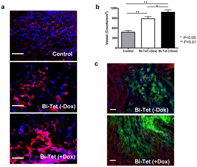
Figure 6. Histology confirming engraftment of transplanted cells.
(a) Representative anti-vWF stainings from animals transplanted with Bi-Tet (+Dox) cells, Bi-Tet (−Dox) cells, and saline control at day 21. (b) Quantitative analysis of capillary density shows improved angiogenesis in the Bi-Tet (+Dox) group compared to the Bi-Tet (−Dox) (*P<0.05) and saline control group (**P<0.01). (c) Immunostaining with troponin-T (red) and GFP (green) demonstrate surviving engrafted cells can incorporate into the host myocardium. Double-positive cells can be found as isolated pockets of cellular islands within the graft as well as in the boundary of transplantation. Nuclei are identified by DAPI staining (blue). Scale bar=50μm.

