
Figure 7. Long-term follow-up of transplanted beating embryoid bodies reveals teratoma formation.
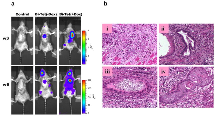
(a) Representative bioluminescence imaging shows teratoma formation in both the Bi-Tet (−Dox) and Bi-Tet (+Dox) animals. (b) Representative postmortem histology of the explanted hearts shows (i) apoptosis of some transplanted cells in the BiTet (−Dox) group as well as differentiation into (ii) glandular epithelia (endoderm), (iii) cartilage (mesoderm), and (iv) squamous epithelia (ectoderm) in the heart.