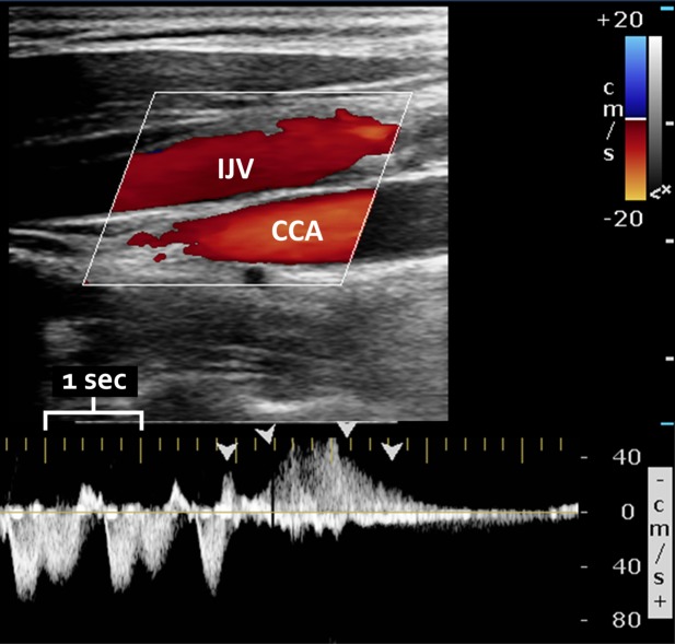
FIGURE 1.

Sample color (top) and spectral (bottom) Doppler ultrasound of the internal jugular vein (IJV), demonstrating reflux (with the same direction of flow as the underlying common carotid artery [CCA]). The large yellow tick marks indicate 1-second intervals. Arrowheads demonstrate reflux lasting >0.88 seconds. Abnormal reflux was only determined using the spectral Doppler waveform, which allows precise determination of flow direction and duration.
