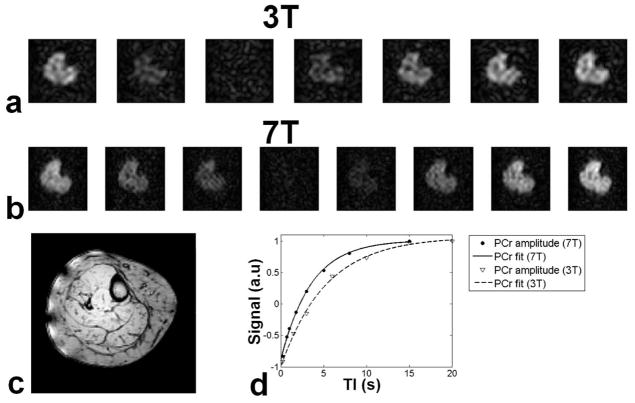
Fig. 3.
Apparent T1 measurement of the same volunteer at 3T and 7T. a) At 3T seven images were acquired at different inversion times TI: 200, 1500, 3000, 6000, 10000 15000, 20000 ms. b) At 7T eight images were acquired at different TI: 200, 700, 1000, 1800, 3000, 5000, 8000, 15000 ms. c) Anatomical image acquired at 7T. d) T1 fitting in the tibialis anterior (TA). At 3T apparent T1 was 5.82 s, which reduced to 3.61 s at 7T.

