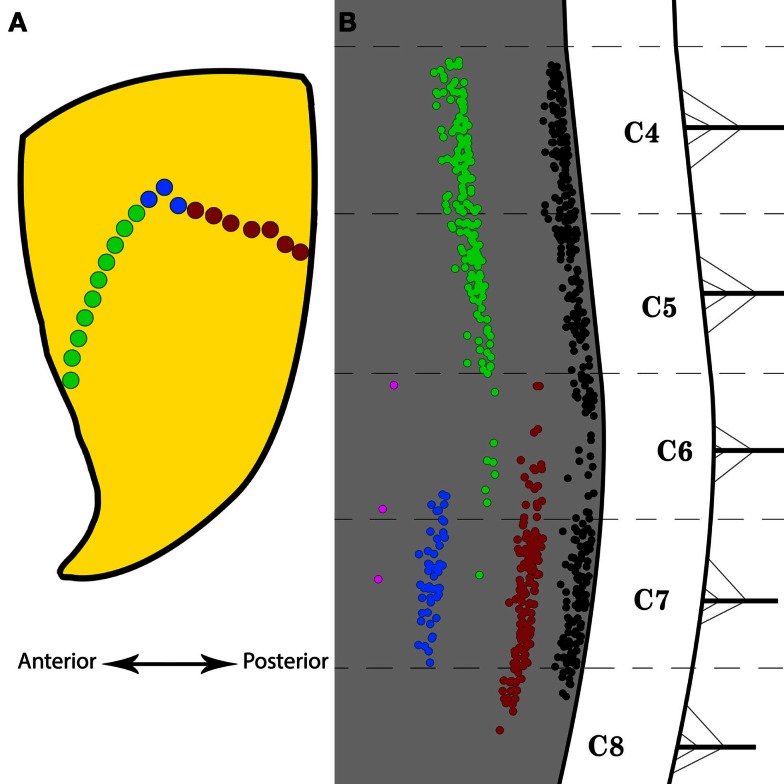
Figure 4.
Selective Fluoro-Gold (FG) targeting of the motor end plate region in triceps brachii and the resulting labeling in the spinal motor neurons. (A) Schematic representation of the motor end plates (MEPs) selectively targeted on the triceps brachii muscle. The green and red dots represent the anterior and posterior halves of the entire MEP region, respectively. The blue dots indicate the location of a bolus injection of FG in the belly of the muscle. The double-headed arrow indicates the antero-posterior direction. (B) Distribution of labeled motor neurons resulting from selective MEP injections of FG as indicated in (A). The black motor neuron column is taken from Figure 3E and represents the typical labeling observed after full-length MEP injections in triceps brachii. The red motor neuron column was obtained after FG injections along the posterior half of the MEP region. The green motor neuron column was obtained after FG injections along the anterior half of the MEP region. The blue motor neuron column was obtained after FG bolus injections in the belly of triceps brachii. The magenta “column” was obtained after application of FG onto the external surface of the fascia over triceps brachii. Each cervical/thoracic spinal cord segment is demarcated by dashed lines. These lines correspond to the halfway point between two nerve roots.

